BLOGGER TEMPLATES AND TWITTER BACKGROUNDS

March 30, 2010

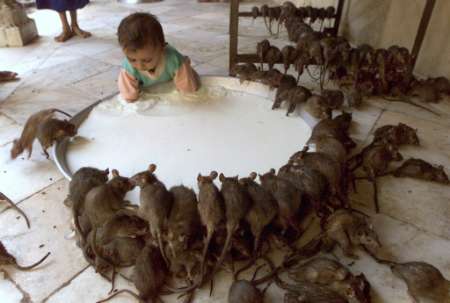
PENYAKIT LEPTOSPIROSIS(kencing tikus)....adakah ini yang menyerang Din Beramboi????


PENYAKIT LEPTOSPIROSIS


Leptospirosis adalah penyakit yang jarang berlaku. Satu penyakit yang tenat/teruk dan boleh berjangkit. Jangkitan berlaku disebabkan oleh beberapa spesies dari keturunan leptospira berbentuk lingkaran (spiral shape).
Nama-nama lain.
Penyakit Weil, Demam Icterohemorrhage, Penyakit Swineherd's, Demam pesawah (Rice-field fever), Demam Pemotong tebu (Cane-cutter fever), Demam Swamp, Demam Lumpur, Jaundis berdarah, Penyakit Stuttgart, Demam Canicola.
Leptospirosis terjadi disebabkan oleh pendedahan kepada kuman yang terdapat di dalam air yang tercemar dengan kencing binatang. Biasanya terjadi di negara beriklim panas

Faktor risiko termasuklah:
Melakukan pekerjaan yang terdedah kepada panas- Petani, penternak/peladang, pekerja rumah penyembelihan, doktor haiwan, pemotong kayu, pekerja sistem pembentungan, pesawah dan anggota tentera.
Aktiviti rekreasi dalam air seperti berenang , berkayak dan berbasikal di laluan denai di kawasan panas.
Terdedah kepada peralatan di rumah termasuk, binatang peliharaan seperti anjing, binatang peliharaan untuk disembelih, ruang perangkap air dan mengambil makanan yang telah digigit oleh binatang seperti tikus, tupai dan arnab.

Leptospirosis ialah penyakit berjangkit disebabkan bakteria Leptospira iaitu bakteria yang berbentuk berpilin (bergelung) yang disebarkan melalui haiwan. Jika tidak dirawat segera, penyakit ini boleh merebak dan membawa maut.

Banyak jenis haiwan boleh menjadi perumah bakteria ini, sama ada liar atau peliharaan. Haiwan dijangkiti ada kalanya menunjukkan tanda sakit, tetapi ada sekadar menjadi pembawa dan tidak mengalami masalah. Secara umumnya bakteria Leptospira boleh menjangkiti  anjing, tikus dan haiwan liar lain seperti rakun dan serigala.


Walaupun catatan kes leptospirosis di negara ini rendah (hanya beberapa kes setahun), ia sebenarnya penyakit global dan kebanyakan kes berlaku di negara beriklim sederhana dan tropika. Di Amerika Syarikat, dianggarkan 100 kes leptospirosis dicatatkan setiap tahun.

Gejala leptospirosis biasanya muncul dalam masa dua hingga 25 hari selepas seseorang itu terdedah kepada kencing, darah atau cebisan tisu haiwan dijangkiti.

Bagaimanapun, bakteria daripada kencing haiwan ini boleh tersebar melalui air atau tanah.

Manusia boleh mendapat jangkitan apabila mengharung atau berenang dalam air tercemar yang tidak mengandungi klorin. Bahkan, menyentuh pokok atau tanah lembap yang tercemar juga boleh memindahkan bakteria kepada manusia selain sentuhan langsung dengan air kencing, darah atau tisu haiwan yang dijangkiti.

Bakteria ini boleh memasuki badan manusia melalui luka atau tisu lembut seperti dalam mulut, hidung atau mata. Bagaimanapun, bakteria ini tidak dipindahkan dari satu individu ke individu lain.
Tanda dan Gejala
Gejala :
Masa pengeraman di antara 2 hingga 26 hari (anggaran 10 hari).
Bermula dengan demam, menggigil, sakit anggota tubuh dan sakit kepala. Gejala ini melibatkan lebih kurang 75% hingga 100% pesakit.
Batuk tidak berkahak (25%-35% kes)
Loya, muntah dan cirit-birit ( 50 kes) Sakit sendi, sakit tulang, sakit tekak dan sakit perut adalah gejala yang kurang berlaku.
Secara umumnya leptospirosis berkembang dalam tiga fasa. Fasa pertama menyebabkan gejala seperti:
Sakit kepala.
Sakit otot.
Mata menjadi sensitif terhadap cahaya terang atau sakit mata.
Seram sejuk.
Demam panas secara tiba-tiba.

Gejala fasa kedua:
Gejala fasa pertama berulang dan demam.
Leher terasa sakit dan tegang.
Sesetengah pesakit mengalami keradangan saraf mata dan selaput otak (meningitis).

Gejala fasa ketiga:
Demam.
Sakit otot.
Jangkitan pada pundi kencing, paru-paru dan jantung (jarang berlaku).

Tanda dan ujian makmal

Radang selaput mata.
Antara 7% hingga 40% pesakit mungkin mengalami sakit otot, pembesaran limpa atau hati, bengkak salur kelenjar, sakit tekak,kekerasan otot, perubahan bunyi pada paru-paru atau ruam.
Dianggarkan 25 hingga 35 peratus pesakit mungkin mengalami batuk tidak berkahak, manakala 50 peratus lagi mempunyai masalah mual, muntah dan cirit-birit. Kebanyakan pesakit (75 hingga 100 peratus) mengalami sakit kepala dan demam.

Pada peringkat seterusnya jangkitan boleh menyebabkan kelenjar limfa bengkak, sakit tekak, otot tegang, bunyi paru-paru yang tidak normal atau ruam. Gejala ini boleh berlanjutan dari beberapa hari ke beberapa minggu.

Dalam kes serius dan tidak mendapat rawatan, leptospirosis boleh menyebabkan kematian. Tahap jangkitan berbeza antara individu mengikut daya ketahanan masing-masing. Ada yang mungkin mengalami gejala teruk, tetapi ada juga individu dijangkiti hanya mendapat gejala ringan.

Masalah kesihatan akibat jangkitan biasanya bermula dengan demam panas secara tiba-tiba.

Gejala menjadi lebih serius apabila penyakit memasuki fasa kedua dan ketiga kerana ia boleh menyebabkan kegagalan buah pinggang dan kerosakan hati.

Ujian sel darah putih (WBC).
Analisa air kencing juga menunjukkan keputusan luar biasa.
Peningkatan kadar creatine kinase dalam darah dikesan pada lebih kurang 50% pesakit.
Antara 40% pesakit mengalami sedikit peningkatan enzim hati.
Diagnosa biasanya dibuat melalui ujian serologi (antibodi).
Kuman dapat dilihat dengan jelas menerusi mikroskop dark field gelap (dark field microscopy), silver stained atau mikroskopi floresent.
Tidak seperti Treponema, pallidum, leptospira boleh membiak melalui darah dan air tulang belakang. Biasanya ia membiak secara perlahan dan pengesanan makmal diperlukan.
50% bilangan kes berjaya mengasingkan kuman dari darah.
Ujian air kencing biasanya positif pada minggu kedua selepas serangan penyakit (illness) dan kekal sehingga 30 hari.

Harapan(Expectations)(prognosis) 
Sembuh dalam kebanyakan kes, bagaimanapun kematian boleh berlaku pada kes yang rumit dan tidak menerima rawatan yang sewajarnya. 


Komplikasi:
  Tindakbalas Jarisch-Herxheimer bila diberi penicillin.
  Jangkitan kuman di otak (Meningitis).
  Perdarahan yang teruk.
  Respiratory distress).
  Kegagalan fungsi hati.
  Kegagalan fungsi buah pinggang 
Rawatan dan Pencegahan
Ubatan Cephalosporim, Penicillins, doxycycline, chloramphenicol, dan erythromycin boleh diberi untuk rawatan leptospirosis. Penjagaan sokongan diperlukan bagi kes yang rumit dan bermasalah.
Apabila dijangkiti, biasanya bakteria Leptospira mengambil masa 10 hari (tempoh inkubasi) sebelum menimbulkan gejala. Namun tempoh inkubasi ini boleh ada dalam julat dua hingga 21 hari. Dalam tempoh ini rawatan sewajarnya perlu diberikan untuk mengelak komplikasi tidak diingini.

Sebagaimana jangkitan bakteria lain, leptospirosis boleh dirawat dengan mengambil antibiotik seperti penisilin, streptomisin, eritromisin, tetrasiklin dan doksisiklin yang perlu diberikan pada peringkat awal jangkitan.

Namun jika gejala dialami serius, doktor akan memantau keadaan pesakit supaya tidak bertambah teruk.

Selain rawatan, beberapa cara boleh dilakukan untuk mengelak jangkitan bakteria leptospira iaitu:


Elak mengharung air (terutama air banjir) atau berenang dalam air sungai atau kolam yang dikhuatiri tercemar, terutama jika anda ada luka atau kudis.


Elak mengunjungi tempat yang dikatakan ada jangkitan buat sementara waktu sehingga ia disahkan selamat.


Jika berenang dalam kolam atau tasik, pastikan air tidak masuk ke dalam mulut.


Pastikan air yang diminum dimasak sehingga mendidih terlebih dulu.


Pastikan kawasan persekitaran anda sentiasa bersih untuk mencegah kedatangan tikus.


Gunakan sarung tangan apabila membuang bangkai haiwan dan membersihkan najis haiwan peliharaan. Bimbang jika haiwan berkenaan juga dijangkiti bakteria leptospira.


Sentiasa bersihkan kawasan keliling rumah supaya air tidak bertakung


lipids,gallstones


Lipids are the one class of large biological molecules that does not consist of polymers. The compounds called lipids are grouped together because they share one important trait: They have little or no affinity for water. The hydrophobic behavior of lipids is based on their molecular structure. Although they may have some polar bonds associated with oxygen, lipids consist mostly of hydrocarbons. Smaller than true (polymeric) macromolecules, lipids are a highly varied group in both form and function. Lipids include waxes and certain pigments, but we will focus on the most biologically important types of lipids: fats, phospholipids, and steroids.
Fats
Although fats are not polymers, they are large molecules, and they are assembled from smaller molecules by dehydration reactions. A fat is constructed from two kinds of smaller molecules: glycerol and fatty acids.

The synthesis and structure of a fat, or triacylglycerol. The molecular building blocks of a fat are one molecule of glycerol and three molecules of fatty acids. (a) One water molecule is removed for each fatty acid joined to the glycerol. (b) A fat molecule with three identical fatty acid units. The carbons of the fatty acids are arranged zig–zag to suggest the actual orientations of the four single bonds extending from each carbon.

Glycerol is an alcohol with three carbons, each bearing a hydroxyl group. A fatty acid has a long carbon skeleton, usually 16 or 18 carbon atoms in length. At one end of the fatty acid is a carboxyl group, the functional group that gives these molecules the name fatty acid. Attached to the carboxyl group is a long hydrocarbon chain. The nonpolar C–H bonds in the hydrocarbon chains of fatty acids are the reason fats are hydrophobic. Fats separate from water because the water molecules hydrogen–bond to one another and exclude the fats. A common example of this phenomenon is the separation of vegetable oil (a liquid fat) from the aqueous vinegar solution in a bottle of salad dressing.

In making a fat, three fatty acid molecules each join to glycerol by an ester linkage, a bond between a hydroxyl group and a carboxyl group. The resulting fat, also called a triacylglycerol , thus consists of three fatty acids linked to one glycerol molecule. (Still another name for a fat is triglyceride, a word often found in the list of ingredients on packaged foods.) The fatty acids in a fat can be the same, as in Figure 5.11b, or they can be of two or three different kinds.

These terms refer to the structure of the hydrocarbon chains of the fatty acids. If there are no double bonds between carbon atoms composing the chain, then as many hydrogen atoms as possible are bonded to the carbon skeleton. Such a structure is described as being saturated with hydrogen, so the resulting fatty acid is called a saturated fatty acid. An unsaturated fatty acid has one or more double bonds, formed by the removal of hydrogen atoms from the carbon skeleton. The fatty acid will have a kink in its hydrocarbon chain wherever a cis double bond occurs.

A fat made from saturated fatty acids is called a saturated fat. Most animal fats are saturated: The hydrocarbon chains of their fatty acids—the “tails” of the fat molecules—lack double bonds, and the molecules can pack tightly, side by side. Saturated animal fats—such as lard and butter—are solid at room temperature. In contrast, the fats of plants and fishes are generally unsaturated, meaning that they are built of one or more types of unsaturated fatty acids. Usually liquid at room temperature, plant and fish fats are referred to as oils—olive oil and cod liver oil are examples. The kinks where the cis double bonds are located prevent the molecules from packing together closely enough to solidify at room temperature. The phrase “hydrogenated vegetable oils” on food labels means that unsaturated fats have been synthetically converted to saturated fats by adding hydrogen. Peanut butter, margarine, and many other products are hydrogenated to prevent lipids from separating out in liquid (oil) form.
A diet rich in saturated fats is one of several factors that may contribute to the cardiovascular disease known as atherosclerosis. In this condition, deposits called plaques develop within the walls of blood vessels, causing inward bulges that impede blood flow and reduce the resilience of the vessels. Recent studies have shown that the process of hydrogenating vegetable oils produces not only saturated fats but also unsaturated fats with trans double bonds. These trans fat molecules may contribute more than saturated fats to atherosclerosis and other problems.

Fat has come to have such a negative connotation in our culture that you might wonder whether fats serve any useful purpose. The major function of fats is energy storage. The hydrocarbon chains of fats are similar to gasoline molecules and just as rich in energy. A gram of fat stores more than twice as much energy as a gram of a polysaccharide, such as starch. Because plants are relatively immobile, they can function with bulky energy storage in the form of starch. (Vegetable oils are generally obtained from seeds, where more compact storage is an asset to the plant.) Animals, however, must carry their energy stores with them, so there is an advantage to having a more compact reservoir of fuel—fat. Humans and other mammals stock their long–term food reserves in adipose cells (see Figure 4.6b), which swell and shrink as fat is deposited and withdrawn from storage. In addition to storing energy, adipose tissue also cushions such vital organs as the kidneys, and a layer of fat beneath the skin insulates the body. This subcutaneous layer is especially thick in whales, seals, and most other marine mammals, protecting them from cold ocean water.


The structure of a phospholipid. A phospholipid has a hydrophilic (polar) head and two hydrophobic (nonpolar) tails. Phospholipid diversity is based on differences in the two fatty acids and in the groups attached to the phosphate group of the head. This particular phospholipid, called a phosphatidylcholine, has an attached choline group. The kink in one of its tails is due to a cis double bond. (a) The structural formula follows a common chemical convention of omitting the carbons and attached hydrogens of the hydrocarbon tails. (b) In the space–filling model, black = carbon, gray = hydrogen, red = oxygen, yellow = phosphorus, and blue = nitrogen.

A phospholipid is similar to a fat, but has only two fatty acids attached to glycerol rather than three. The third hydroxyl group of glycerol is joined to a phosphate group, which has a negative electrical charge. Additional small molecules, usually charged or polar, can be linked to the phosphate group to form a variety of phospholipids.

Phospholipids show ambivalent behavior toward water. Their hydrocarbon tails are hydrophobic and are excluded from water. However, the phosphate group and its attachments form a hydrophilic head that has an affinity for water. When phospholipids are added to water, they self–assemble into double–layered aggregates—bilayers—that shield their hydrophobic portions from water.

Steroids
Steroids are lipids characterized by a carbon skeleton consisting of four fused rings.

Different steroids vary in the functional groups attached to this ensemble of rings. One steroid, cholesterol , is a common component of animal cell membranes and is also the precursor from which other steroids are synthesized. Many hormones, including vertebrate sex hormones, are steroids produced from cholesterol. Thus, cholesterol is a crucial molecule in animals, although a high level of it in the blood may contribute to atherosclerosis. Both saturated fats and trans fats exert their negative impact on health by affecting cholesterol levels.

What are gallstones?
Gallstones are small, pebble-like substances that develop in the gallbladder. The gallbladder is a small, pear-shaped sac located below your liver in the right upper abdomen. Gallstones form when liquid stored in the gallbladder hardens into pieces of stone-like material. The liquid—called bile—helps the body digest fats. Bile is made in the liver, then stored in the gallbladder until the body needs it. The gallbladder contracts and pushes the bile into a tube—called the common bile duct—that carries it to the small intestine, where it helps with digestion.

Bile contains water, cholesterol, fats, bile salts, proteins, and bilirubin—a waste product. Bile salts break up fat, and bilirubin gives bile and stool a yellowish-brown color. If the liquid bile contains too much cholesterol, bile salts, or bilirubin, it can harden into gallstones.

The two types of gallstones are cholesterol stones and pigment stones. Cholesterol stones are usually yellow-green and are made primarily of hardened cholesterol. They account for about 80 percent of gallstones. Pigment stones are small, dark stones made of bilirubin. Gallstones can be as small as a grain of sand or as large as a golf ball. The gallbladder can develop just one large stone, hundreds of tiny stones, or a combination of the two.

Gallstones can block the normal flow of bile if they move from the gallbladder and lodge in any of the ducts that carry bile from the liver to the small intestine. The ducts include the hepatic ducts, which carry bile out of the liver cystic duct, which takes bile to and from the gallbladder common bile duct, which takes bile from the cystic and hepatic ducts to the small intestine.

Bile trapped in these ducts can cause inflammation in the gallbladder, the ducts, or in rare cases, the liver. Other ducts open into the common bile duct, including the pancreatic duct, which carries digestive enzymes out of the pancreas. Sometimes gallstones passing through the common bile duct provoke inflammation in the pancreas—called gallstone pancreatitis—an extremely painful and potentially dangerous condition.


If any of the bile ducts remain blocked for a significant period of time, severe damage or infection can occur in the gallbladder, liver, or pancreas. Left untreated, the condition can be fatal. Warning signs of a serious problem are fever, jaundice, and persistent pain.

What causes gallstones?
Scientists believe cholesterol stones form when bile contains too much cholesterol, too much bilirubin, or not enough bile salts, or when the gallbladder does not empty completely or often enough. The reason these imbalances occur is not known.

The cause of pigment stones is not fully understood. The stones tend to develop in people who have liver cirrhosis, biliary tract infections, or hereditary blood disorders—such as sickle cell anaemia—in which the liver makes too much bilirubin.

The mere presence of gallstones may cause more gallstones to develop. Other factors that contribute to the formation of gallstones, particularly cholesterol stones, include

Sex. Women are twice as likely as men to develop gallstones. Excess estrogen from pregnancy, hormone replacement therapy, and birth control pills appears to increase cholesterol levels in bile and decrease gallbladder movement, which can lead to gallstones.

Family history. Gallstones often run in families, pointing to a possible genetic link.

Weight. A large clinical study showed that being even moderately overweight increases the risk for developing gallstones. The most likely reason is that the amount of bile salts in bile is reduced, resulting in more cholesterol. Increased cholesterol reduces gallbladder emptying. Obesity is a major risk factor for gallstones, especially in women.

Diet. Diets high in fat and cholesterol and low in fiber increase the risk of gallstones due to increased cholesterol in the bile and reduced gallbladder emptying.

Rapid weight loss. As the body metabolizes fat during prolonged fasting and rapid weight loss—such as “crash diets”—the liver secretes extra cholesterol into bile, which can cause gallstones. In addition, the gallbladder does not empty properly.

Age. People older than age 60 are more likely to develop gallstones than younger people. As people age, the body tends to secrete more cholesterol into bile.

Ethnicity. American Indians have a genetic predisposition to secrete high levels of cholesterol in bile. In fact, they have the highest rate of gallstones in the United States. The majority of American Indian men have gallstones by age 60.

Cholesterol-lowering drugs. Drugs that lower cholesterol levels in the blood actually increase the amount of cholesterol secreted into bile. In turn, the risk of gallstones increases.

Diabetes. People with diabetes generally have high levels of fatty acids called triglycerides. These fatty acids may increase the risk of gallstones.

Who is at risk for gallstones?

People at risk for gallstones include :
women—especially women who are pregnant, use hormone replacement therapy, or take birth control pills
people over age 60
overweight or obese men and women
people who fast or lose a lot of weight quickly
people with a family history of gallstones
people with diabetes
people who take cholesterol-lowering drugs

What are the symptoms of gallstones?
As gallstones move into the bile ducts and create blockage, pressure increases in the gallbladder and one or more symptoms may occur. Symptoms of blocked bile ducts are often called a gallbladder “attack” because they occur suddenly. Gallbladder attacks often follow fatty meals, and they may occur during the night. A typical attack can causesteady pain in the right upper abdomen that increases rapidly and lasts from 30 minutes to several hours pain in the back between the shoulder blades pain under the right shoulder

Notify your doctor if you think you have experienced a gallbladder attack. Although these attacks often pass as gallstones move, your gallbladder can become infected and rupture if a blockage remains.

People with any of the following symptoms should see a doctor immediately:
prolonged pain—more than 5 hours
nausea and vomiting
fever—even low-grade—or chills
yellowish color of the skin or whites of the eyes
clay-coloured stools

Many people with gallstones have no symptoms; these gallstones are called “silent stones.” They do not interfere with gallbladder, liver, or pancreas function and do not need treatment.

How are gallstones diagnosed?
Frequently, gallstones are discovered during tests for other health conditions. When gallstones are suspected to be the cause of symptoms, the doctor is likely to do an ultrasound exam—the most sensitive and specific test for gallstones. A handheld device, which a technician glides over the abdomen, sends sound waves toward the gallbladder. The sound waves bounce off the gallbladder, liver, and other organs, and their echoes make electrical impulses that create a picture of the gallbladder on a video monitor. If gallstones are present, the sound waves will bounce off them, too, showing their location. Other tests may also be performed.

Computerized tomography (CT) scan. The CT scan is a noninvasive x ray that produces cross-section images of the body. The test may show the gallstones or complications, such as infection and rupture of the gallbladder or bile ducts.

Cholescintigraphy (HIDA scan). The patient is injected with a small amount of nonharmful radioactive material that is absorbed by the gallbladder, which is then stimulated to contract. The test is used to diagnose abnormal contraction of the gallbladder or obstruction of the bile ducts.

Endoscopic retrograde cholangiopancreatography (ERCP). ERCP is used to locate and remove stones in the bile ducts. After lightly sedating you, the doctor inserts an endoscope—a long, flexible, lighted tube with a camera—down the throat and through the stomach and into the small intestine. The endoscope is connected to a computer and video monitor. The doctor guides the endoscope and injects a special dye that helps the bile ducts appear better on the monitor. The endoscope helps the doctor locate the affected bile duct and the gallstone. The stone is captured in a tiny basket and removed with the endoscope.

Blood tests. Blood tests may be performed to look for signs of infection, obstruction, pancreatitis, or jaundice.

Because gallstone symptoms may be similar to those of a heart attack, appendicitis, ulcers, irritable bowel syndrome, hiatal hernia, pancreatitis, and hepatitis, an accurate diagnosis is important.

How are gallstones treated?
Surgery. If you have gallstones without symptoms, you do not require treatment. If you are having frequent gallbladder attacks, your doctor will likely recommend you have your gallbladder removed—an operation called a cholecystectomy. Surgery to remove the gallbladder—a nonessential organ—is one of the most common surgeries performed on adults in the United States.

Nearly all cholecystectomies are performed with laparoscopy. After giving you medication to sedate you, the surgeon makes several tiny incisions in the abdomen and inserts a laparoscope and a miniature video camera. The camera sends a magnified image from inside the body to a video monitor, giving the surgeon a close-up view of the organs and tissues. While watching the monitor, the surgeon uses the instruments to carefully separate the gallbladder from the liver, bile ducts, and other structures. Then the surgeon cuts the cystic duct and removes the gallbladder through one of the small incisions.

Recovery after laparoscopic surgery usually involves only one night in the hospital, and normal activity can be resumed after a few days at home. Because the abdominal muscles are not cut during laparoscopic surgery, patients have less pain and fewer complications than after “open” surgery, which requires a 5- to 8-inch incision across the abdomen.

If tests show the gallbladder has severe inflammation, infection, or scarring from other operations, the surgeon may perform open surgery to remove the gallbladder. In some cases, open surgery is planned; however, sometimes these problems are discovered during the laparoscopy and the surgeon must make a larger incision. Recovery from open surgery usually requires 3 to 5 days in the hospital and several weeks at home. Open surgery is necessary in about 5 percent of gallbladder operations.

The most common complication in gallbladder surgery is injury to the bile ducts. An injured common bile duct can leak bile and cause a painful and potentially dangerous infection. Mild injuries can sometimes be treated nonsurgically. Major injury, however, is more serious and requires additional surgery.

If gallstones are present in the bile ducts, the physician—usually a gastroenterologist—may use ERCP to locate and remove them before or during gallbladder surgery. Occasionally, a person who has had a cholecystectomy is diagnosed with a gallstone in the bile ducts weeks, months, or even years after the surgery. The ERCP procedure is usually successful in removing the stone in these cases.

Nonsurgical Treatment.Nonsurgical approaches are used only in special situations—such as when a patient has a serious medical condition preventing surgery—and only for cholesterol stones. Stones commonly recur within 5 years in patients treated nonsurgically.

Oral dissolution therapy. Drugs made from bile acid are used to dissolve gallstones. The drugs ursodiol (Actigall) and chenodiol (Chenix) work best for small cholesterol stones. Months or years of treatment may be necessary before all the stones dissolve. Both drugs may cause mild diarrhea, and chenodiol may temporarily raise levels of blood cholesterol and the liver enzyme transaminase.

Contact dissolution therapy. This experimental procedure involves injecting a drug directly into the gallbladder to dissolve cholesterol stones. The drug—methyl tert-butyl ether—can dissolve some stones in 1 to 3 days, but it causes irritation and some complications have been reported. The procedure is being tested in symptomatic patients with small stones.

Do people need their gallbladder?
Fortunately, the gallbladder is an organ people can live without. Your liver produces enough bile to digest a normal diet. Once the gallbladder is removed, bile flows out of the liver through the hepatic ducts into the common bile duct and directly into the small intestine, instead of being stored in the gallbladder. Because now the bile flows into the small intestine more often, softer and more frequent stools can occur in about 1 percent of people. These changes are usually temporary, but talk with your health care provider if they persist.

Points to Remember
Gallstones form when bile hardens in the gallbladder.

Gallstones are more common among older adults; women;  people with diabetes; those with a family history of gallstones; people who are overweight, obese, or undergo rapid weight loss; and those taking cholesterol-lowering drugs.

Gallbladder attacks often occur after eating a meal, especially one high in fat.

Symptoms can mimic those of other problems, including a heart attack, so an accurate diagnosis is important.

Gallstones can cause serious problems if they become trapped in the bile ducts.

Laparoscopic surgery to remove the gallbladder is the most common treatment.

March 27, 2010

Penggodam,Penganggur,Penceroboh....


GAMBAR menunjukkan penggodam Twitter milik Obama yang dikenali sebagai Francois C


CLERMONT-FERRAND – Seorang penggodam komputer di Perancis berdepan hukuman penjara semalam selepas dia memecah masuk akaun laman jaringan sosial Twitter milik Presiden Amerika Syarikat (AS), Barack Obama dan selebriti Britney Spears dengan meneka kata laluan mereka.


Penggodam berusia 23 tahun yang cuma dikenali sebagai Francois C. menggunakan nama anjing kesayangan Obama, Bo sebagai kata laluan akaun Twitter milik presiden AS itu.
Biro Penyiasatan Persekutuan (FBI) dengan bantuan polis Perancis mengambil masa enam bulan menjejaki lelaki itu yang menganggur sebelum menangkapnya di sini, tengah Perancis pada Selasa lepas.
“Saya tidak berniat jahat… Saya cuma mahu memberi amaran kepada mereka bahawa terdapat kelemahan dalam sistem yang digunakan,” kata lelaki itu.
Penggodam tersebut yang berdepan hukuman penjara dua tahun sekiranya didapati bersalah ditahan di sebuah balai polis di sini sebelum dibebaskan dengan ikat jamin bagi menunggu perbicaraan pada 24 Jun depan. – Agensi
kredit : kosmo.com.my :twisted:

Kenapa mengantuk lepas makan?


MENGANTUKNYA! Setiap kali setelah makan tengah hari mesti mengantuk. Kadangkala rasa tidak larat hendak buka mata. Rasanya bukan karena tidak cukup tidur sebab tiap-tiap malam aku tidur nyenyak, menggerutu seorang teman yang beberapa kali tersengguk-sengguk depan komputer.
Sememangnya, mengantuk setelah makan bukan karena kurang tidur, sebaliknya ia menandakan kita dahaga dan badan tidak cukup air. Ramai tersilap menafsirkan kebutuhan tubuh terhadap air dengan rasa lapar.
Dahaga bukan petanda terbaik untuk memberitahu badan memerlukan air karena sensitivitas terhadap dahaga semakin berkurang apabila umur meningkat, terutama pada orang tua. Badan kita hanya dapat mengatur keseimbangan air sampai satu tingkat tertentu.

Paras garam akan meningkat apabila penyimpanan air dalam badan berkurang. Otak akan mendeteksi perubahan keseimbangan ini dan memberi isyarat kepada buah pinggang untuk mengurangi kehilangan air dan memicu rasa dahaga.
Bagaimanapun, cetusan rasa dahaga ini terjadi lewat. Biasanya apabila kita merasa dahaga, badan sudah kehilangan kira-kira dua cangkir cairan tubuh. Kehilangan ini dapat menimbulkan berbagai implikasi.
Air adalah unsur penting dalam badan. Badan lelaki yang melibatkan 65 persen air dan 55 persen bagi wanita. Air bertindak sebagai pelarut, penyejuk, pelumas dan agen pengangkut nutrien ke seluruh badan.
Kandungan air dalam tubuh setiap orang adalah berbeda menurut kandungan lemak masing-masing. Secara umumnya, jumlah persentase air dalam tubuh orang yang tidak banyak lemak adalah lebih tinggi. Kira-kira 70 persen berat otak, 75 persen berat jantung, 90 persen berat paru-paru dan 82 persen kandungan darah melibatkan air.
Badan mengalami kehilangan air sepanjang masa, hampir 80 persen sehari, walaupun kita tidak aktif. Kehilangan ini terjadi melalui air kencing, najis, peluh dan pernafasan. Setiap kali menghembuskan nafas, kita kehilangan air dalam bentuk uap. Jika dikumpulkan, kita mungkin bisa dapat satu atau dua gelas air yang hilang melalui pernafasan.
Ini sebabnya pakar kesehatan menyarankan kita minum sekurang-kurangnya delapan gelas atau satu liter air setiap hari. Sumber air bukan hanya dari air minum karena ia juga ada dalam makanan, buah-buahan dan sayur-sayuran.
Berapa banyak air yang kita perlukan?
Secara umumnya setiap orang membutuhkan sekurang-kurangnya delapan gelas air atau satu liter air sehari. Namun, jumlah ini berbeda antara setiap individu, aktivitas dilakukan dan cuaca tempat tinggal. Tentu saja jika kita melakukan latihan pada hari panas, kita kehilangan lebih banyak air dalam peluh.
Air yang keluar ini perlu diganti segera, sama ada ketika atau setelah berolahraga, untuk menghindari masalah kekeringan. Kehilangan banyak air menyebabkan tekanan darah turun mendadak, suplai oksigen ke otak menurun dan jika terlalu serius, ia boleh membawa maut.
Jika terlalu banyak minum, apakah akan terjadi keracunan air? Memang boleh, tetapi ia jarang terjadi. Sebenarnya sulit bagi orang sehat mengalami keracunan air. Ia biasanya berlaku pada bayi. Malah, individu yang alami masalah berat badan berlebihan harus minum lebih banyak air, yaitu segelas untuk setiap 11 kilogram melebihi berat badan ideal.
Mereka yang bercita-cita mengurangi berat badan juga dianjurkan minum banyak air. Kekurangan air merendahkan kadar metabolisme dan badan tidak boleh memecah dan menggunakan lemak dengan efektif. Akibatnya, proses menurunkan berat badan menjadi lebih sulit.
Masalah retensi air atau TIDAK badan mengeluarkan air dari pundi kencing menjadi lebih buruk jika kita tidak mendapat cukup air. Ada kalanya masalah ini dapat diatasi dengan minum lebih banyak air untuk menggalakkan pembuangan air berlebihan.
Oleh karena sebagian besar zat makanan larut dalam air, mungkin ada yang risau ia dibuang melalui air kencing. Jangan khawatir. Masalah ini tidak berlaku kerana buah pinggang kita memiliki sistem penyaringan yang cerdik. Hanya bahan tidak berguna dibuang. Semua khasiat makanan diserap ketika dalam usus dan dimasukkan ke dalam darah untuk didistribusikan ke seluruh badan.
Sebenarnya banyak mengalami masalah kekurangan air tanpa disadari. Malah, banyak juga mengatakan tidak mau kerap berulang ke toilet sebagai alasan untuk tidak minum banyak air. Tindakan ini sebenarnya meracun diri karena tanpa air yang cukup, badan sulit mengeluarkan bahan toksik dan dapat berkumpul membentuk batu karang.
Tanpa air yang cukup juga, nutrisi dari makanan tidak dapat didistribusikan dengan baik dan menyebabkan lebih banyak lemak terkumpul dalam badan. Bentuk dan ukuran otot turut berubah. Paling malang, jika kekurangan air meningkatkan tingkat bahan toksik hingga membahayakan nyawa.
Kesan kekurangan cairan
Kajian menunjukkan kehilangan dua persen cairan tubuh bisa menyebabkan:
Lemah daya ingatan (ingatan jangka pendek)
Sukar membuat perkiraan, walaupun yang Mudah
Sukar memberi tumpuan terhadap perkara dilakukan terutama pada layar komputer atau bahan bacaan tercetak
Tekanan darah menurun
Jumlah oksigen yang sampai ke otak berkurang menyebabkan rasa mengantuk
Lesu pada waktu siang
Sakit punggung
Dehidrasi
Sakit kepala
Memperlahankan kadar metabolisme
Meningkatkan risiko kanker usus, pundi kencing dan payu dara
Sendi dan otot mengalami kekeringan
Menyukarkan penyingkiran bahan beracun
Meningkatkan keinginan untuk makan
Darah menjadi pekat
Mudah keliru.
Detak jantung meningkat
Mempengaruhi reaksi kimia dalam badan